Describe the Structure of the Retina
It consists of lens retina optic nerve aqueous humor and vitreous humor. The conversion of electromagnetic entry into nerve impulses is achieved by the.
Layers Of The Retina Discovery Eye Foundation
Retina is the innermost layer.
. MLO 143 Describe the structure of the nose and the pathway of olfactory information. MLO 141 Describe the structure and function of the eye and accessory eye structures MLO 142 Define how light is focused on the retina leading to phototransduction then how this information is relayed into the brain. The retina is structured of several layers of nerve cells photoreceptors pigmentation and blood vessels which line the inside of the eyeball.
The high density of cones in the macula makes the visual image detailed just as a high-resolution digital camera has more megapixels. It is the outer covering a protective tough white layer called the sclera white part of the eye. Describe the structure location and function of the fovea centralis central hole in macula lutea area of highest visual acuity contains high density of cones reason for eye movement.
Want to see the full answer. C The innermost nervous coat of eye contains retina. The receptor cells present in the retina are of two types rod cells and cone cells.
The retina is the sensory membrane that lines the inner surface of the back of the eyeball. See also Chapter 22. It consists of the following parts.
Describe the structure of the retina and the cells that compose it. The front transparent part of the sclera is called cornea. The eye is the sensory organ that perceives the sense of vision and has structural features which can receive light and convert them into neural impulses.
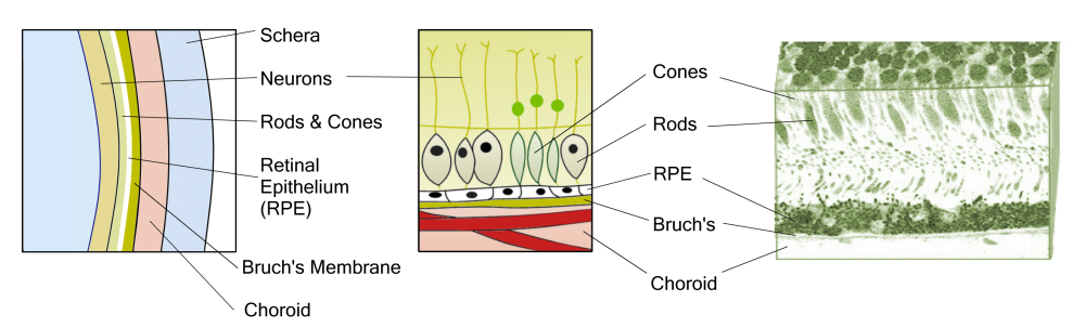
The retina is most closely linked with the underlying layers of the eyeball along the edge of the optic nerve head. The outermost layer closest to the sclera consists of pigment cells. The central portion of the retina is called the.
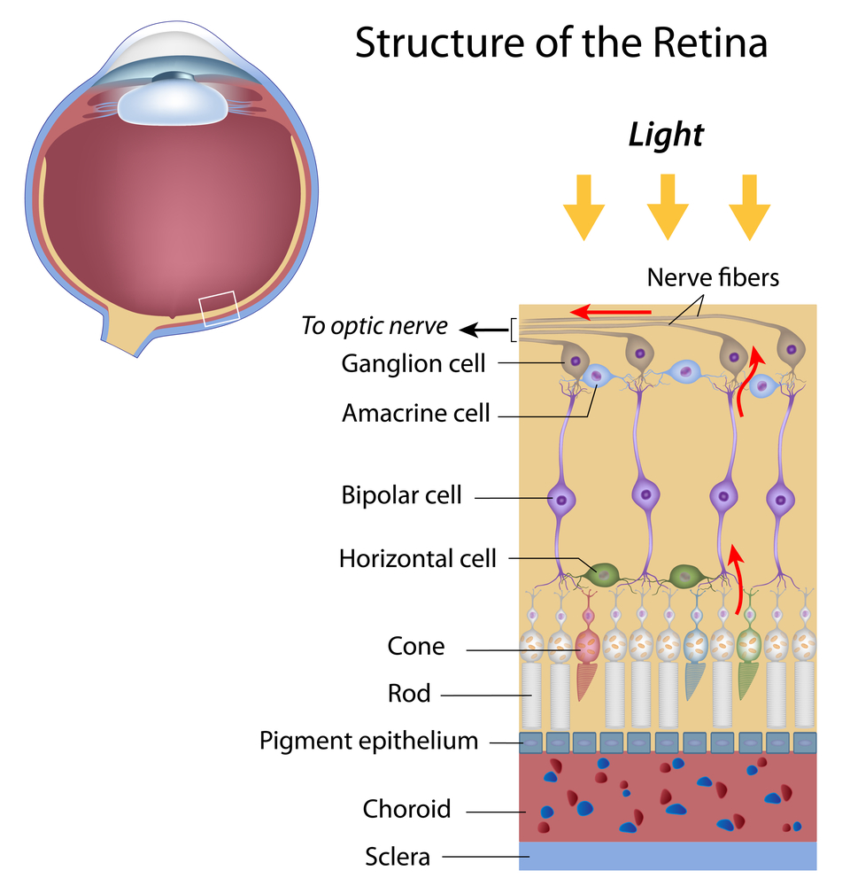
During development the retina forms as an outpocketing of the diencephalon called the optic vesicle which undergoes invagination to form the optic cup Figure 113. It contains three layers of cells inner ganglion cells middle bipolar cells and outermost photoreceptor cells. The receptor cells present in the retina are of two types rod cells and cone cells.
Solution for Describe the structure of the retina. Describe the structure of the retina and how light affects rhodopsin. The outer nuclear layer ONL composed of the cell bodies of the rods and cones is about the same thickness in central and peripheral retina.
The retina is the screen at the end of the eye where all the images are formed. The inner wall of the optic cup gives rise to the retina while the outer wall gives. It is the innermost layer of the eye.
Each layer of cells in this tissue serves a specific purpose. The lens along with the cornea refracts the entering light and helps to focus it on the retina. The retina spreads around the inside of the entire back of the eye.
Despite its peripheral location the retina or neural portion of the eye is actually part of the central nervous system. Approximately 125 million rods and 1 million cones are present in each human eye. The retina is the innermost layer.
The internal structure of the eye is a little complex. The most sensitive part of the retina is a small area called the macula which has millions of tightly packed photoreceptors the type called cones. To focus visual input to this spot.
The purpose of the retina is to receive light that the lens has focused. Terms in this set 6 What structure absorbs stray light and prevents reflection. Its composed of several layers including one that contains specialized cells called photoreceptors.
Step 1 of 3. The retina is a light-sensitive layer of nerve tissue lining the inner surface of the eye. It is extremely sensitive to light because of the presence of Photoreceptors which are photosensitive cells that detect dim and colored lights.
The retina creates an image projected on its surface with help of the cornea and crystalline lens and transforms it into nerve impulses sent to the brain. Structure of the retina. Light enters the eye through the cornea.
The retina at the back of the eye is essential for all vision. The internal structure of the eye includes retina lens aqueous humor vitreous humor and optic nerve. There are two types of photoreceptor cells in the human eye rods and cones.
Adjacent to this layer are the photoreceptors rods and cones. The retina is composed of several cell layers. Three layers of neural cells are present in them they are ganglion bipolar and photoreceptor cells.
It converts the image into electrical. The retina contains the cells that sense light photoreceptors and the blood vessels that nourish them. Central retina is cone-dominated retina whereas peripheral retina is rod-dominated.
It contains three layers of cells inner ganglion cells middle bipolar cells and outermost photoreceptor cells. Thus in central retina the cones are closely spaced and the rods fewer in number between the cones Figs. Describe the structure of the retina and explain how light stimulates action potentials in the optic nerves.
Rod photoreceptors detect motion provide black-and. It is light sensitive and acts as a film of a camera. Biology questions and answers.
The most posterior layer at the very back of the eye Pigment epithelium. The lens is the transparent biconvex structure that is attached to the ciliary body by ligament tissue. It is located near the optic nerve.
The retina is a thin layer of tissue that lines the back of the eye on the inside. Describe the structure of the retina and explain howlight stimulates action potentials in the optic nerves. As we prepare for Age-Related Macular Degeneration Awareness Month in February a closer look at the layers of the retina and their function.
Retina Structure

Human Eye The Retina Britannica

Schematic Of The Eye And Retina Structure The Magnified Area Download Scientific Diagram
No comments for "Describe the Structure of the Retina"
Post a Comment